Systemic lupus erythematosus (SLE) is an autoimmune disease where the host immune system attacks its own tissues, causing widespread inflammation and tissue damage in the affected organs. Increased inflammation affects the joints, skin, brain, lungs, kidneys, and blood vessels.
Patients with SLE may experience a variety of symptoms that include fatigue, skin rashes, fevers, and pain or swelling in the joints. Pathologies of human SLE include lymph node enlargement, proteinuria, and kidney failure. Autoantigens such as anti-nuclear antibodies as well as skin-specific anti-desmoglin 3 (Dsg3) may also be involved in tissue inflammation.
Scientists at Aragen created the SLE model – MRL/lpr lupus female mice and investigated the effects of Methylprednisolone, a typical SLE medication. MLR/lpr mice are homozygous for a spontaneous mutation in the FAS gene and develop lymphadenopathy (enlarged lymph nodes), splenomegaly (enlarged spleen), anti-nuclear and anti-dsDNA antibodies, and systemic autoimmunity. Autoimmune responses occur spontaneously in these mice, resulting in arthritis and glomerulonephritis mimicking SLE symptoms in humans.
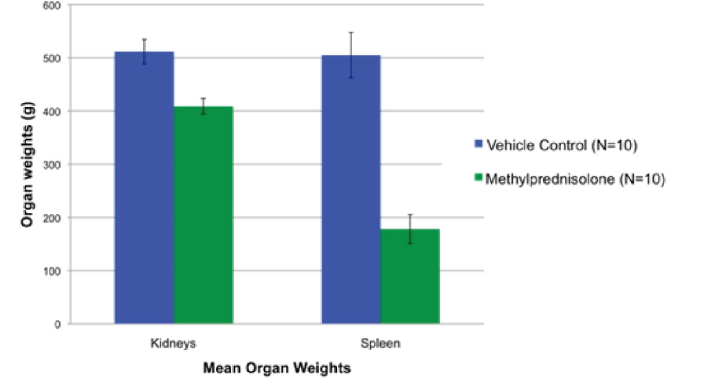
The experiments were conducted in two groups, both of which have MRL/lpr lupus 13-week old female mice. The animals were selected based on their proteinuria scores at 12 weeks of age. Animals that were tested for Methylprednisolone were termed positive control whereas the negative control group was treated with Methylcellulose. The observations were recorded until the mice reached the age of 19 weeks. The effect of the drug was studied by recording the change in the body weight on weekly basis. The urine was tested on a weekly basis for proteinuria scores whereas the serum testing was done every two weeks to check anti-ds DNA antibody levels and anti-Dsg 3 antibody levels. At the end of the 13th week, two organs i.e., spleen and kidney were harvested from both the animals, and their weight measured.
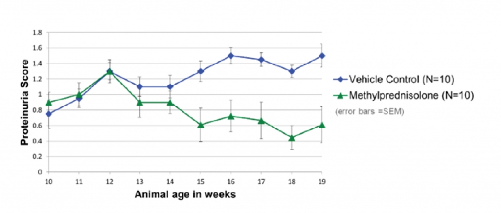
Aragen scientists observed that the methylprednisolone-treated group has lower proteinuria scores compared to the negative control group (Figure 1). Proteinuria scores were higher in SLE patients as compared to healthy individuals.
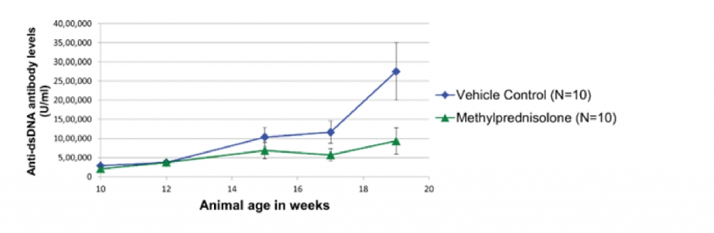
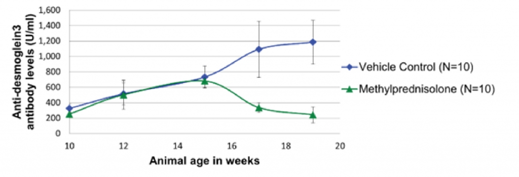
Serum analysis of both the treatment groups shows that the positive control group has lower concentrations of both anti-ds DNA antibody (Figure 2) and anti-Dsg 3 antibody (Figure 3) levels.
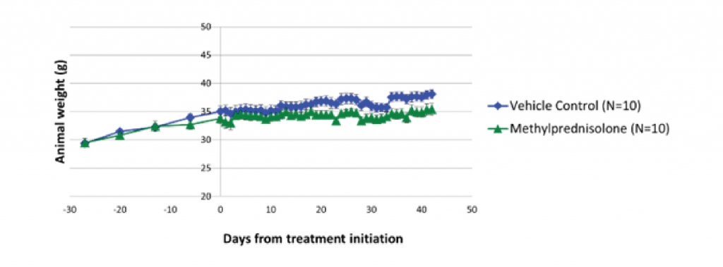
Body weights of both the treatment groups demonstrated that the positive control group has lower body weight after the medication (Figure 4). Moreover, it was observed that the weight of the kidney and spleen decreased in the positive control group as compared to the negative control group (Figure 5).
MRL/lpr mice are a spontaneous model for SLE. These models responded very well to the standard medication i.e., methylprednisolone. These models are suitable for studying the pathophysiology of the SLE and are modeled specifically to conduct preclinical studies of the novel drug compounds against SLE.