

Understanding the durability of weight-loss therapies is critical for developing effective long-term obesity management strategies. Semaglutide has emerged as a highly effective therapy for obesity, delivering substantial weight loss and metabolic benefits through GLP-1 receptor activation. This case study evaluates the effects of semaglutide treatment, withdrawal, and re-treatment on metabolic health, body composition, muscle function, and behavioral outcomes in a diet-induced obesity mouse model.
Although semaglutide has demonstrated robust weight-loss efficacy, the durability of its benefits following treatment discontinuation and the response to therapy re-initiation remain incompletely understood. Furthermore, a comprehensive understanding of how repeated treatment cycles influence metabolic health, body composition, muscle function, behavioral outcomes, and obesity-related comorbidities is needed to inform long-term treatment strategies.
As outlined in the study design (Figure 1), male C57BL/6JHsd mice were fed a 60% kcal high-fat diet for 16 weeks to establish diet-induced obesity (DIO). Following a 5-day acclimation period, animals were randomized based on body weight and treated twice daily with vehicle or semaglutide (30 nmol/kg, SC). The study comprised three phases: an initial treatment phase to evaluate weight-loss and metabolic responses, a treatment withdrawal phase to assess weight regain and metabolic rebound, and a re-treatment phase to evaluate responsiveness following therapy re-initiation.
Throughout the study, longitudinal monitoring of body weight, food intake, water intake, and body composition was conducted to assess treatment-related changes in energy balance and adiposity. Additional assessments included:
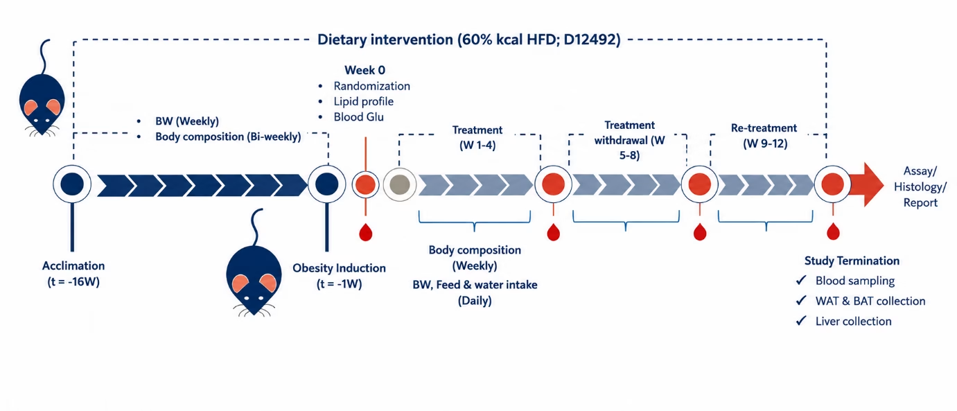
Figure 1: Study design for evaluating the effects of semaglutide treatment, withdrawal, and re-treatment in a diet-induced obesity (DIO) mouse model.
Reproducible Weight Loss Upon Re-treatment
Semaglutide induced substantial weight loss during the initial treatment phase. Following treatment withdrawal, animals rapidly regained weight, indicating reversal of the therapeutic effect. However, re-initiation of semaglutide produced a second robust weight-loss response comparable to that observed during the initial treatment period, demonstrating preserved responsiveness despite prior drug exposure.
Consistent with these findings, semaglutide significantly reduced food intake during both treatment phases. Food consumption increased during the washout period and paralleled body weight regain, while re-treatment restored appetite suppression, confirming sustained pharmacological efficacy (Figure 2).
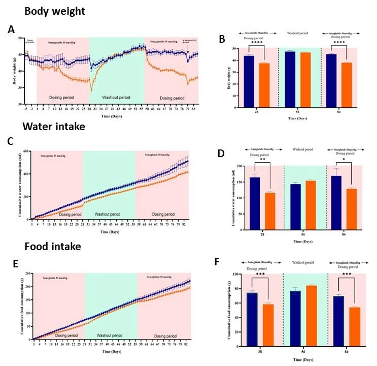
Figure 2: Effects of semaglutide treatment, withdrawal, and re-treatment on body weight, cumulative water intake, and cumulative food intake in diet-induced obese (DIO) mice. (A-B) Body weight. (C-D) Water intake. (E-F) Food intake. Bars represent vehicle-treated DIO mice and orange bars represent semaglutide-treated DIO mice.
Improved Glycemic Control
Semaglutide treatment was also associated with improved glycemic control. Blood glucose levels were significantly reduced at the end of the initial treatment phase (Day 27) compared with vehicle-treated controls and remained lower following re-treatment (Day 84). Although blood glucose levels increased during the washout period, the reduction observed upon re-treatment demonstrates restoration of semaglutide’s glucose-lowering effects, consistent with the weight-loss response (Figure 3).
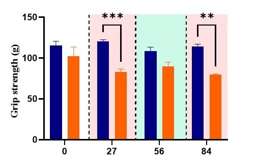
Figure 3: Effects of semaglutide treatment, withdrawal, and re-treatment on blood glucose levels in diet-induced obese (DIO) mice. Blood glucose was measured on Days 0, 27, 56, and 84. Blue bars represent vehicle-treated DIO mice and orange bars represent semaglutide-treated DIO mice.
Changes in Body Composition
Body composition analysis was performed to determine whether semaglutide-induced weight loss was driven primarily by reductions in fat mass or accompanied by changes in lean and fluid mass (Figure 4).
Repeated semaglutide treatment significantly reduced fat mass, with partial recovery observed during the washout phase. Treatment was also associated with modest but significant reductions in lean and fluid mass following re-treatment.
These findings indicate that semaglutide-mediated weight loss was primarily driven by fat loss but was accompanied by changes in lean and fluid mass, highlighting the importance of comprehensive body composition assessments during long-term obesity studies.
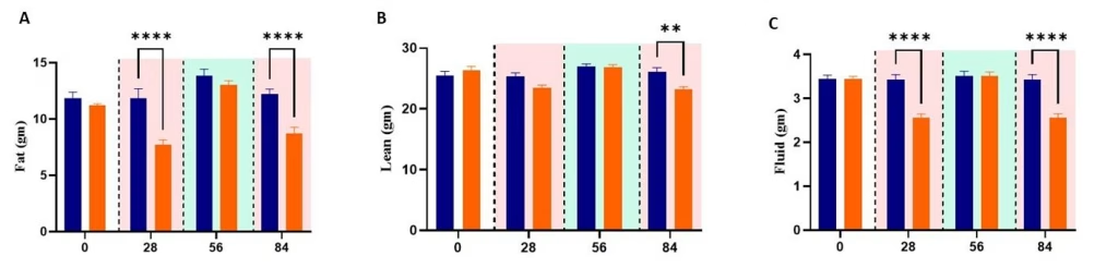
Figure 4: Effects of semaglutide treatment, withdrawal, and re-treatment on body composition in diet-induced obese (DIO) mice. (A) Fat mass, (B) lean mass, and (C) fluid mass measured at baseline (Day 0), end of the treatment phase (Day 28), end of the washout phase (Day 56), and end of the re-treatment phase (Day 84). Blue bars represent vehicle-treated DIO mice and orange bars represent semaglutide-treated DIO mice.
Effects on Muscle Function
To evaluate the functional consequences of repeated semaglutide treatment, grip strength was assessed throughout the study (Figure 5). Semaglutide-treated animals exhibited a significant reduction in grip strength following the initial treatment phase, with a further decline observed after re-treatment. While grip strength partially recovered during the washout period, values remained lower than those of vehicle-treated controls at the end of the study.
These findings suggest that repeated semaglutide treatment may affect muscle performance, highlighting the importance of incorporating functional assessments alongside conventional metabolic endpoints.
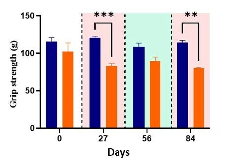
Figure 5: Effects of semaglutide treatment, withdrawal, and re-treatment on grip strength in diet-induced obese (DIO) mice to assess changes in muscle function. Grip strength was measured on Days 0, 28, 56, and 84 to assess changes in muscle function. Blue bars represent vehicle-treated DIO mice and orange bars represent semaglutide-treated DIO mice.
Behavioral Adaptations
Sucrose preference was assessed at the end of the study as a measure of reward-associated feeding behavior and overall physiological well-being (Figure 6).
Semaglutide-treated animals exhibited a significant reduction in sucrose preference compared with vehicle-treated controls, indicating reduced interest in rewarding stimuli. This finding suggests that repeated semaglutide treatment may influence reward-related feeding and potentially induce anhedonia-like behavior in addition to its established effects on appetite and body weight regulation.
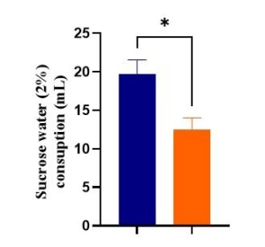
Figure 6: Effects of repeated semaglutide treatment on sucrose preference in diet-induced obese (DIO) mice. Sucrose preference was measured at day 79 to assess treatment-related changes in reward-associated feeding behavior. X-axis represents treatment groups: blue bars represent vehicle-treated DIO mice and orange bars represent semaglutide-treated DIO mice.
Adipose Tissue Remodeling
Histological and morphometric analyses of epididymal white adipose tissue (eWAT) revealed treatment-associated remodeling following semaglutide administration (Figure 7). Representative H&E-stained sections (Figure 7A) showed reduced adipocyte size in semaglutide-treated animals compared with vehicle controls. This observation was further supported by segmented adipocyte images used for morphometric analysis (Figure 7B).
At the tissue level, semaglutide treatment reduced eWAT mass (Figure 7C). In addition, quantitative analysis demonstrated a decrease in adipocyte cross-sectional area (Figure 7D), indicating reduced lipid storage within individual adipocytes.
Together, these findings demonstrate that semaglutide-mediated weight loss was accompanied by favorable adipose tissue remodeling and reduced adiposity.
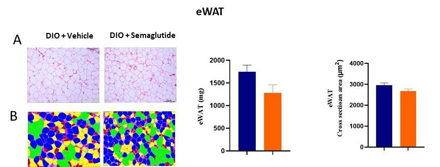
Figure 7: Histological and morphometric evaluation of epididymal white adipose tissue (eWAT) following semaglutide treatment in diet-induced obese (DIO) mice. (A) Representative H&E-stained eWAT sections from vehicle- and semaglutide-treated animals. (B) Representative segmented images used for adipocyte morphometric analysis. (C) Quantification of eWAT mass. (D) Quantification of eWAT adipocyte cross-sectional area. In quantitative graphs, x-axis represents treatment groups: blue bars represent vehicle-treated DIO mice and orange bars represent semaglutide-treated DIO mice.
Perirenal Adipose Tissue Remodeling
Histological and morphometric analyses of perirenal adipose tissue demonstrated remodeling following semaglutide treatment (Figure 8). Representative H&E-stained sections (Figure 8A) showed smaller adipocytes in semaglutide-treated animals compared with vehicle controls. This observation was further supported by segmented images used for adipocyte morphometric analysis (Figure 8B).
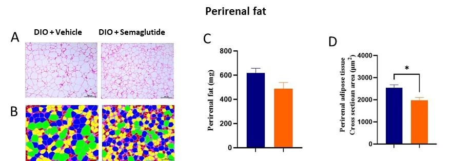
Figure 8: Histological and morphometric evaluation of perirenal adipose tissue following semaglutide treatment in diet-induced obese (DIO) mice. (A) Representative H&E-stained perirenal adipose tissue sections from vehicle- and semaglutide-treated animals. (B) Representative segmented images used for perirenal adipocyte morphometric analysis. (C) Quantification of perirenal fat mass. (D) Quantification of perirenal adipocyte cross-sectional area. In quantitative graphs, x-axis represents treatment groups: blue bars represent vehicle-treated DIO mice and orange bars represent semaglutide-treated DIO mice.
At the tissue level, semaglutide treatment reduced perirenal fat mass (Figure 10C). Quantitative analysis also revealed a significant decrease in adipocyte cross-sectional area (Figure 10D), indicating reduced lipid storage within individual adipocytes.
These findings demonstrate that semaglutide-mediated weight loss was accompanied by remodeling of perirenal adipose tissue and reduced adiposity across multiple fat depots.
Brown Adipose Tissue Remodeling
Histological and morphometric analyses of interscapular brown adipose tissue (iBAT) demonstrated significant tissue remodeling following semaglutide treatment (Figure 9). Representative H&E-stained sections (Figure 9A) revealed smaller brown adipocytes in semaglutide-treated animals compared with vehicle controls, which was further visualized through segmented images used for morphometric analysis (Figure 9B).
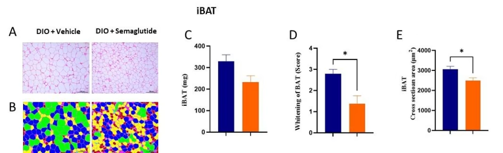
Figure 9: Histological and morphometric evaluation of interscapular brown adipose tissue (iBAT) following semaglutide treatment in diet-induced obese (DIO) mice. (A) Representative H&E-stained iBAT sections from vehicle- and semaglutide-treated animals. (B) Representative segmented images used for iBAT adipocyte morphometric analysis. (C) Quantification of iBAT mass. (D) BAT whitening score. (E) Quantification of iBAT adipocyte cross-sectional area. In quantitative graphs, x-axis represents treatment groups: blue bars represent vehicle-treated DIO mice and orange bars represent semaglutide-treated DIO mice.
At the tissue level, semaglutide treatment reduced iBAT mass (Figure 9C). In addition, a significant reduction in BAT whitening score (Figure 9D) was observed, indicating improved brown adipose tissue morphology and metabolic status. Quantitative analysis also showed a decrease in adipocyte cross-sectional area (Figure 9E), consistent with reduced lipid accumulation within brown adipocytes.
Overall, these findings demonstrate that semaglutide promotes favorable remodeling of brown adipose tissue, characterized by reduced adiposity and improved BAT morphology.
Improved Liver Health
Histopathological evaluation revealed marked improvements in liver health following semaglutide treatment (Figure 10). Representative Oil Red O-stained liver sections (Figure 10A) demonstrated reduced hepatic lipid accumulation in semaglutide-treated animals compared with vehicle controls. Consistent with these findings, H&E-stained liver sections (Figure 10B) showed a substantial reduction in steatotic lesions and improved liver morphology.
Quantitative analyses further confirmed these observations. Semaglutide treatment significantly reduced liver weight (Figure 10C), decreased the percentage of lipid-stained liver area (Figure 10D), and improved overall liver histology scores (Figure 10E), indicating attenuation of obesity-associated hepatic steatosis.
Collectively, these findings demonstrate that semaglutide not only promotes weight loss but also improves liver pathology associated with obesity.
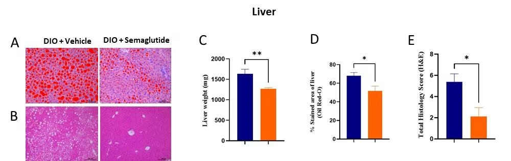
Figure 10: Histopathological assessment of liver steatosis following semaglutide treatment in diet-induced obese (DIO) mice. (A) Representative Oil Red O-stained liver sections showing hepatic lipid accumulation. (B) Representative H&E-stained liver sections showing liver morphology and steatotic changes. (C) Quantification of liver weight. (D) Percentage Oil Red O-stained liver area. (E) Total liver histology score based on H&E evaluation. In quantitative graphs, x-axis represents treatment groups: blue bars represent vehicle-treated DIO mice and orange bars represent semaglutide-treated DIO mice.
Effects on Muscle Function
Repeated semaglutide treatment was associated with reductions in lean body mass and significantly lower grip strength, suggesting an impact on muscle performance. To further investigate these effects, quadriceps muscle morphology and fiber characteristics were evaluated (Figure 11).
Histological assessment of quadriceps muscle (Figure 11A) showed no overt structural abnormalities between treatment groups. Semaglutide-treated animals exhibited a significant reduction in quadriceps muscle mass (Figure 11C); however, segmented muscle fiber analysis (Figure 11B) and quantification of skeletal muscle fiber cross-sectional area (Figure 11D) revealed no significant differences in muscle fiber size.
These findings indicate that while semaglutide treatment reduced muscle mass and functional performance, skeletal muscle architecture remained largely preserved.
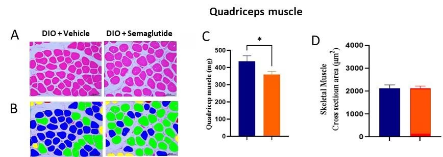
Figure 11: Histological and morphometric evaluation of quadriceps muscle following semaglutide treatment in diet-induced obese (DIO) mice. (A) Representative H&E-stained quadriceps muscle sections from vehicle- and semaglutide-treated animals. (B) Representative segmented images used for skeletal muscle fiber morphometric analysis. (C) Quantification of quadriceps muscle mass. (D) Quantification of skeletal muscle fiber cross-sectional area. In quantitative graphs, x-axis represents treatment groups: blue bars represent vehicle-treated DIO mice and orange bars represent semaglutide-treated DIO mice.
Semaglutide delivered robust and reproducible weight loss, with efficacy restored upon re-treatment following withdrawal. While treatment discontinuation led to rapid weight regain, re-initiation effectively reinstated metabolic benefits, including improved glycemic control, reduced adiposity, and improved hepatic steatosis.
Repeated treatment also revealed reductions in lean mass, grip strength, and sucrose preference, suggesting potential effects on muscle function and physiological well-being, despite largely preserved skeletal muscle morphology. Using a clinically relevant DIO model and integrated metabolic, functional, behavioral, histological, and morphometric endpoints, this study provided a comprehensive assessment of treatment durability, tissue-level adaptations, and the broader physiological effects of long-term obesity therapy.
Developing differentiated obesity therapies requires a deeper understanding of treatment durability, metabolic adaptations, and physiological outcomes beyond weight loss alone. Aragen’s integrated obesity research platform combines translationally relevant models, innovative study designs, and multidimensional endpoint assessments to generate actionable insights that support critical development decisions. Our capabilities include:
By integrating metabolic, functional, behavioral, and histopathological assessments within a single study framework, Aragen provides a comprehensive view of efficacy, durability, and safety, accelerating the development of next-generation obesity and metabolic disease therapies.
Ready to Advance Your Obesity Research?
Partner with Aragen to leverage robust obesity models, translational endpoints, and integrated preclinical expertise to evaluate the efficacy, durability, and safety of next-generation metabolic therapies.
